
☰
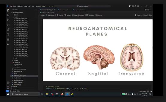
Advanced deep learning solution for precise 3D segmentation of brain tumors from MRI scans, enabling accurate glioma identification, therapy planning, and early detection through neuroanatomical plane analysis.
The project focuses on the management of Gliomas, which are recognized as highly aggressive brain tumors that present formidable challenges within the fields of neurology and oncology.
Effective management of these tumors necessitates high-precision identification, meticulous therapy planning, and timely detection to significantly enhance patient survival rates and clinical outcomes. Currently, the primary diagnostic tool is Magnetic Resonance Imaging (MRI), which provides detailed structural brain data essential for tumor analysis.
The implementation of advanced 3D segmentation algorithms aims to transform healthcare by improving the accuracy, efficiency, and consistency of glioma management through deep learning and multi-modal MRI analysis.
The core challenge in the current clinical workflow is the heavy reliance on manual segmentation of MRI scans by medical professionals. This manual approach is critically flawed as it is extremely labor-intensive and highly susceptible to human error, which can lead to inconsistent diagnostic results.
Without precise automated boundaries, clinicians struggle to achieve the level of accuracy required for optimal pathological analysis and early-stage detection. There is a profound need for robust, automated 3D segmentation tools that can complete, multi-modal MRI data to provide reliable, scalable, and standardized results that support clinical decision-making.
To address these challenges, the project proposes a comprehensive AI-driven system that integrates state-of-the-art deep learning with enterprise-grade engineering: